Next generation ultrasound imaging research is focused on a new sensor technology which utilizes large arrays of small membrane sensors. Accurate performance from these sensors can enable a new suite of medical imaging applications, allowing doctors to more rapidly test and diagnose their patients.
The capacitive micromachined ultrasonic transducer (CMUT) is an emerging ultrasound sensor technology that may replace piezoelectric sensors. A CMUT is a vacuum-sealed membrane, which can be electrostatically actuated to send and receive acoustic signals. These membranes can be fabricated in large quantities using standard microfabrication procedures, allowing for the creation of smaller transducer elements to be used for higher resolution imaging.
CMUT technology offers several advantages over conventional piezoelectric sensors. They offer a larger signal bandwidth with adjustable target frequencies dependent on the physical size of each membrane. CMUTs are also compatible with CMOS circuitry allowing for integrated driving electronics on the chip, further reducing any noise from the system.
Larger sensor arrays often utilize tens of thousands or more CMUT cells to provide larger imaging fields of view. After fabricating these sensors, it is important to evaluate their operation to guarantee uniform performance across the entire array for accurate medical imaging applications. Our group uses laser Doppler vibrometry to non-destructively evaluate the resonant frequency and membrane displacement of our sensors prior to packaging.
Our system
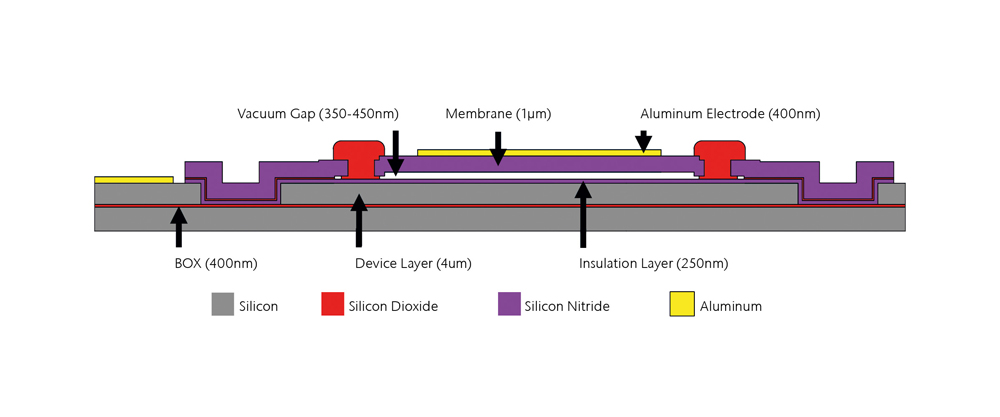
Our CMUT cells are fabricated using two layers of sacrificial poly-silicon patterned to form the vacuum gap. The membranes are deposited on top of the sacrificial layers and are composed of a low-stress silicon nitride film that is released using a heated potassium hydroxide solution to dissolve the poly-silicon films. Subsequently, the volume formally occupied by the sacrificial layers is sealed under vacuum using silicon dioxide plugs. A cross-section of an individual CMUT membrane is shown in figure 1.
In order to operate the large number of CMUT cells required for a greater field-of-view, our CMUT sensors are fabricated in square arrays with rows and columns connected to groups of membranes. Each intersection between the top and bottom orthogonal electrodes forms a single imaging element. For our quality control measurements, we evaluate each CMUT array in an open air medium.
Measuring device performance
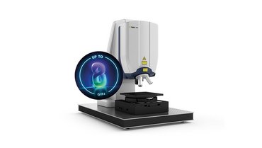
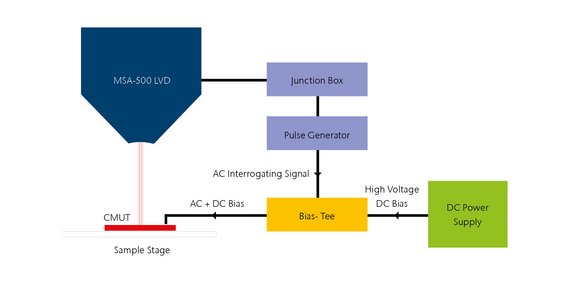
For our characterization measurements, we utilize Polytec’s MSA Micro System Analyzer Laser Doppler Vibrometer. Each CMUT array is mounted on a sample stage and connected using three-axis micropositioning probes. Each array is driven using a combined AC and DC bias voltage connected using a bias-tee as shown in figure 2.
When evaluating the performance of a sensor array, we focus on measurements of the resonant frequency and dynamic displacements for different AC and DC driving conditions. We are interested in comparing results between different positions across the device, to analyze the general performance of the array, and to investigate any displacement changes due to resistive losses and to spot any defective membranes.
Evaluation and future plans
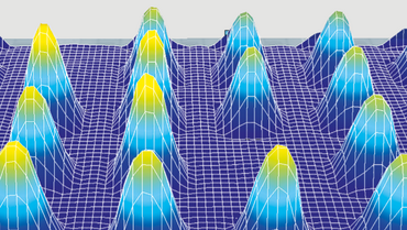
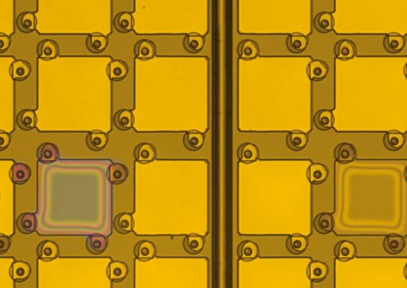
When evaluating CMUT cells, it is critical that every membrane operates identically, otherwise small deviations across the array, or clusters of dead cells, will create artifacts while imaging with these sensors. Some of these variations can be spotted visually using a microscope, such as collapsed or deformed cells shown in figure 3.
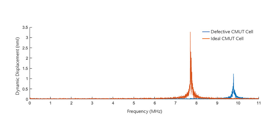
However, if the sealing plugs leak in any of the CMUTs, then the cavities will fill with air causing a shift in the resonant frequency and performance of the cell, while appearing through the microscope to be no different from a vacuum sealed membrane. This shift in resonant frequency, as well as a decrease in dynamic displacement, is caused by the damping effects of squeezing and stretching a pocket of air at the Megahertz frequencies used for medical imaging. These defects normally can only be observed when operating the sensor, so it is beneficial to probe the device and determine if these defects exist before spending additional resources connecting and packaging the sensor for testing in an ultrasound system.
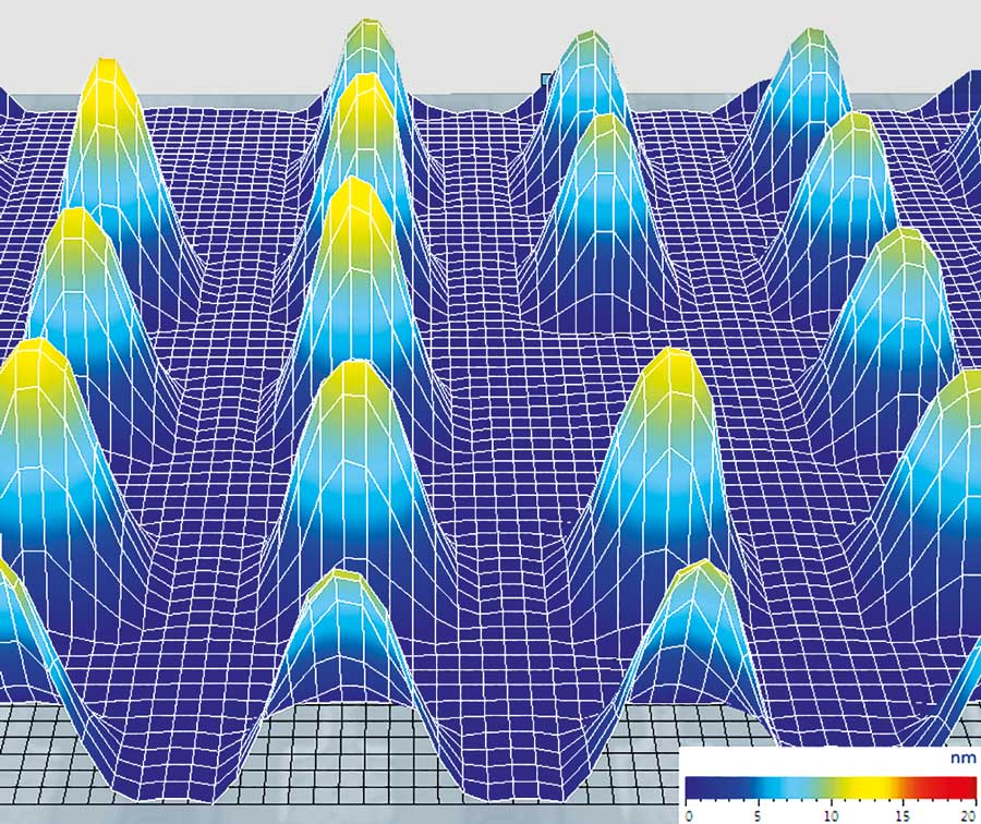
An example of several leaking membranes is shown in figure 4a, where 2 cells are not resonating at the same frequency as the rest of the array when operating with sinewave excitation. We can further investigate the operating frequency of both the ideal CMUT cell and the defective sensor using a pseudorandom excitation signal with the vibrometer, as shown in figure 4b. As you can see the air present in the membrane shifts the resonant frequency higher while also decreasing the dynamic displace ment for a given operating voltage.
Successful design and fabrication of CMUT medical imaging transducers is dependent on a refined process flow to produce defect free products. It is useful in this development process to have equipment capable of inspecting each array to ensure proper quality control standards are being met. Using Polytec’s MSA Laser Doppler Vibrometer we are able to inspect our sensors during the fabrication process so that we can identify and correct production errors, and avoid spending additional resources packaging and testing sensors we already know to be defective.
Images courtesy: Images courtesy of the authors unless otherwise specified. Cover image: ©istockphoto.com/cjspencer


